Reading, Technology and the Brain
“Scientists now use PET scans and other advanced imaging techniques to study the living, working brain. Their efforts are giving us fascinating glimpses into how the brain works during reading, and provide insight into how a balanced approach to the process of learning to read should be designed.”- Teaching Every Student in the Digital Age (Ch 1)
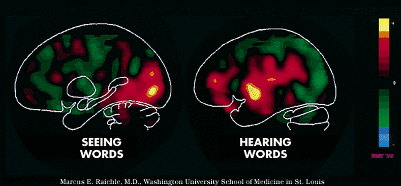
PET scans show that there are several different parts of the brain that are involved in reading, each making its own contribution to success.
PET scans generate images of the brain that distinguish highly active regions from those that are less active. The more active a region is, the more glucose it metabolizes — creating a “hot spot” of energy consumption. The greater the activity, the more intense the hot spot, and the more brightly colored its appearance on the PET scan.
Seeing Words
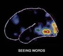
Seeing words mainly involves areas in the occipital lobes. The dramatic differences in brain function during these two activities begin to suggest how complex learning to read is. The scans also imply something else. The active parts of the brain work together to perform what may superficially seem a simple task.
Hearing Words
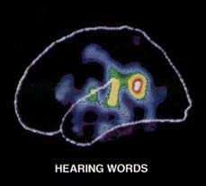
Hearing words creates hot spots mainly in the temporal lobe of the cortex.
The scan below is a view of both sides of the brain. The PET scan on the left shows two areas of the brain (red and yellow) that become particularly active when volunteers read words on a video screen:the primary visual cortex and an additional part of the visual system, both in the back of the left hemisphere. Other brain regions become especially active when subjects hear words through ear-phones, as seen in the PET scan on the right.
The scan below is a view of both sides of the brain. The PET scan on the left shows two areas of the brain (red and yellow) that become particularly active when volunteers read words on a video screen:the primary visual cortex and an additional part of the visual system, both in the back of the left hemisphere. Other brain regions become especially active when subjects hear words through ear-phones, as seen in the PET scan on the right.